
Jawbone Cavitations & NICO Treatment Melbourne
Assessment and biological surgical management of suspected jawbone cavitations, NICO lesions and areas of compromised jawbone healing.
Jawbone cavitations are a term sometimes used to describe areas of altered or compromised bone healing within the jaw, often associated with previous extraction sites such as wisdom teeth and root canal-treated teeth.
In scientific and dental literature, these findings have been described using several terms, including NICO lesions (neuralgia-inducing cavitational osteonecrosis), osteonecrosis and fatty degenerative osteonecrosis of the jaw (FDOJ) and residual bone cysts. Importantly, the terminology, diagnostic criteria and clinical significance of these findings continue to evolve and remain debated within the fields of dentistry and oral medicine.
At Laser and Holistic Dental in Melbourne, we provide assessment of suspected jawbone cavitations and NICO lesions using comprehensive clinical examination and, where clinically indicated, 3D CBCT imaging to evaluate previous extraction sites and surrounding jawbone in three dimensions.
Where surgical treatment is considered clinically appropriate, our biological oral surgery approach may incorporate carefully selected techniques to remove compromised tissue and manage the surgical site, followed by biocompatible bone grafting and regenerative materials intended to support new bone formation and remodelling within the treated bony defect.
Book a Cavitation & Jawbone Assessment →
What Is a Jawbone Cavitation or NICO Lesion?
Following the removal of a tooth, the extraction site normally undergoes a process of blood clot formation, tissue repair and new bone formation.
In some patients, an extraction site may demonstrate areas of incomplete or altered bone healing. These findings are particularly discussed in relation to previous wisdom tooth extraction sites and root canal filled teeth – although they may occur elsewhere within the jaws.
NICO (neuralgia-inducing cavitational osteonecrosis) is terminology that has historically been used in the scientific literature to describe areas of jawbone osteonecrosis associated particularly with facial or neuralgic pain. Other terminology encountered in the literature includes jawbone cavitation, ischemic osteonecrosis and fatty degenerative osteonecrosis of the jaw (FDOJ) and residual bone cysts.
When a diseased or infected tooth is removed, our biological approach includes careful management of the extraction site and removal of diseased or compromised tissue, including periodontal ligament and inflammatory tissue where clinically indicated. The surrounding bone is also assessed and managed according to the individual site. Areas of compromised bone or residual pathological tissue within a previous extraction site may be considered when investigating suspected jawbone cavitations or NICO lesions.
These terms are not necessarily interchangeable, and there is currently no universally accepted diagnostic standard for NICO. Not every area of altered bone healing or radiographic irregularity represents NICO or requires surgical treatment.
How Are Jawbone Cavitations & NICO Lesions Assessed?
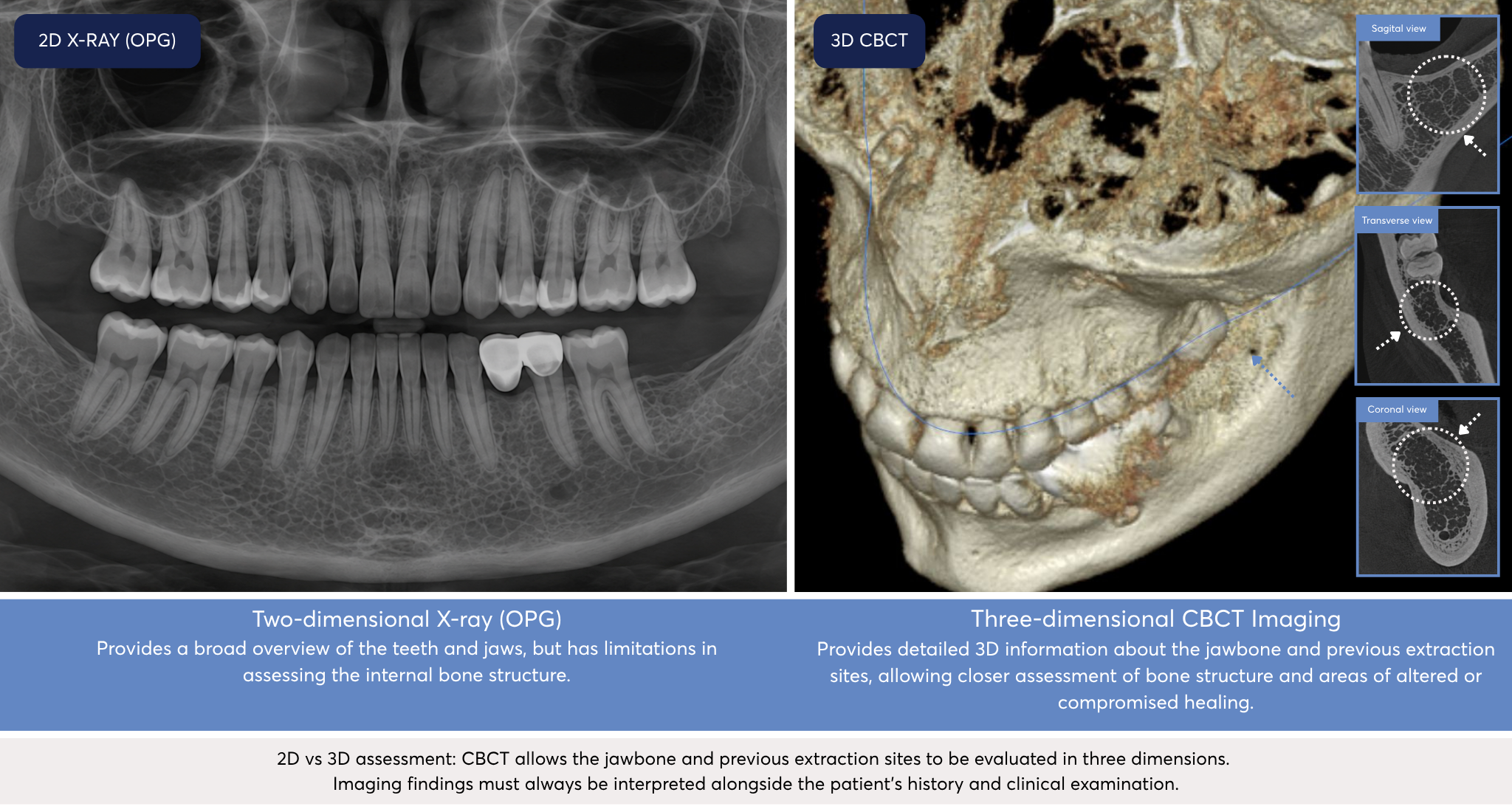
Two-dimensional dental X-rays provide useful information but can have limitations when assessing the internal architecture of the jawbone.
3D CBCT for Jawbone Cavitation Assessment
- For patients where there is a clinical reason to investigate a previous extraction or surgical site, 3D Cone Beam Computed Tomography (CBCT) can provide significantly more anatomical information about the area.
Cavitations & NICO After Wisdom Tooth Removal
Previous wisdom tooth extraction sites are one of the areas that may be investigated when assessing suspected jawbone cavitations or NICO lesions.
Where a patient has persistent concerns around a previous wisdom tooth site, assessment may include examination of the area and review of previous dental imaging. When clinically justified, 3D CBCT imaging can provide additional information about the bone architecture, healing pattern and surrounding anatomical structures.
An abnormal appearance on imaging does not by itself establish a diagnosis of NICO or mean that surgery is required. Findings must be considered alongside the patient’s clinical history, examination and symptoms.
Our Cavitation Assessment & Treatment Journey
1. 3D CBCT & Comprehensive Consultation
-
- Assessment begins with a detailed dental and medical history, clinical examination and, where clinically indicated, 3D CBCT imaging and referral for blood tests.
- The CBCT allows our dentists to examine previous extraction sites and surrounding anatomical structures in three dimensions and identify areas that may warrant further investigation.
- Your dentist will review the findings with you and discuss whether monitoring, further investigation, referral or treatment may be appropriate.
Explore 3D CBCT Imaging →
2. Biological Cavitation Surgery & NICO Curettage
-
- Where cavitation surgery or NICO curettage is considered clinically appropriate, treatment generally involves accessing the affected area and carefully removing necrotic or compromised bone and tissue.
- Depending upon the individual case, our biological surgical protocol may incorporate:
- Precision surgical and piezoelectric instrumentation
- Er:YAG and/or Nd:YAG dental laser technology
- Medical-grade ozone
- Autologous Concentrated Growth Factors (CGF) prepared from the patient’s own blood
- Carefully selected biocompatible bone-grafting materials and local anaesthetic
- Techniques designed to support the surgical environment and normal healing
These technologies and regenerative materials are incorporated as part of our carefully developed biological surgical approach, with treatment individually planned to support the surgical environment, bone healing and remodelling.
The exact surgical protocol varies according to the patient’s anatomy, medical history, clinical findings and treatment requirements.
Learn more about our approach to eliminating oral infection →
3. Post-Operative Laser & Healing Support
-
- Healing is monitored through scheduled post-surgical reviews as part of our ongoing regenerative approach.
- Post-operative care may incorporate photobiomodulation and laser therapy, including treatment directed towards the surgical area and surrounding jaw, TMJ and lymphatic regions. These therapies form part of our broader post-surgical approach designed to support and accelerate the healing response following surgery.
- Follow-up care allows our dentists to monitor healing, manage post-operative symptoms and identify any areas requiring further assessment or treatment.
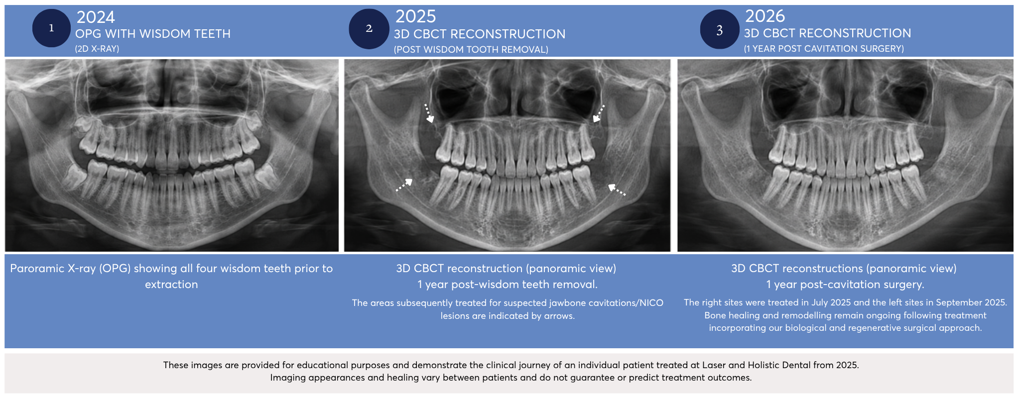
Before, During & After Cavitation Treatment
The clinical journey below demonstrates the assessment, surgical treatment and ongoing healing of an individual patient following our biological and regenerative approach to cavitation surgery.
Symptoms & Reasons Patients Seek NICO or Cavitation Assessment
Patients may seek assessment of a previous extraction site for a variety of reasons. Some experience persistent or unexplained jaw or facial discomfort, while others have been advised that imaging may demonstrate an area requiring further assessment or have specifically been referred for assessment of a suspected NICO lesion or jawbone cavitation.
A consultation may be considered where there is:
- Persistent pain or tenderness around a previous extraction site
- Unexplained jaw or facial pain
- Altered sensation around a previous surgical area
- A history of complicated healing or infection following an extraction
- An area of altered bone identified during dental imaging
- Concern regarding a previous cavitation procedure that has not healed as expected
Importantly, these symptoms can have many different causes and do not necessarily indicate the presence of a cavitation. A comprehensive assessment is required before any diagnosis or treatment recommendation is made.
Revision Cavitation & NICO Surgery
Previously Had Cavitation or NICO Surgery?
Laser and Holistic Dental also assesses patients who have undergone previous cavitation surgery, NICO surgery or jawbone curettage elsewhere and continue to have clinical concerns.
Our biological dentists can review your previous treatment history, existing imaging and current clinical presentation and, where appropriate, undertake new 3D imaging to assess the area.
If further treatment is clinically indicated, your dentist will discuss the available options and whether revision cavitation surgery may be appropriate.
Book a Second Opinion or Cavitation Assessment →
Travelling to Melbourne for Cavitation Treatment
Interstate & Overseas Cavitation Patients
Laser and Holistic Dental regularly welcomes patients travelling to Melbourne from interstate and overseas for jawbone cavitation and NICO assessment, alongside other complex biological dental treatments.
Our team can help coordinate your consultation, CBCT imaging, surgery and appropriate post-operative reviews to reduce unnecessary travel where clinically possible.
If you have existing X-rays, CBCT scans or dental records, these can be provided to our team before your visit for review.
For patients who have undergone previous cavitation or NICO treatment elsewhere, we also offer assessment of persistent or recurrent areas of concern.
Interstate & Overseas Patient Guide →
Why patients choose a holistic dentist →
Dr Dome & Biological Dentistry
Dr Dominik Nischwitz (Dr Dome) has extensively discussed jawbone cavitations, including what he describes as FDOK (Fatty Degenerative Osteonecrosis of the Jawbone), within his approach to biological dentistry. Dr Dome has also spoken publicly about his own personal experience undergoing cavitation surgery, providing him with both a professional and personal perspective on this area of dentistry. His clinical approach considers previous extraction sites, particularly wisdom tooth sites, and incorporates 3D imaging alongside biological surgical techniques including piezo surgery, ozone and autologous blood-derived materials.
At Laser and Holistic Dental, we similarly take a comprehensive biological approach when assessing areas of previous extraction and suspected jawbone cavitations. Our biological dentists provide assessment and treatment planning for patients concerned about jawbone cavitations and NICO lesions, using the patient’s clinical history, examination and appropriate imaging, including 3D CBCT where clinically indicated.
Because terminology, diagnostic criteria and the clinical significance of NICO/FDOK remain areas of ongoing scientific discussion, imaging findings are considered as part of the overall clinical assessment rather than in isolation.
Explore Dr Dome’s approach to FDOK, NICO & cavitations →
Understanding NICO, Jawbone Cavitations & the Scientific Evidence
NICO (neuralgia-inducing cavitational osteonecrosis) and jawbone cavitations have been described in dental and medical literature for several decades. Other terminology encountered in published research includes osteonecrosis, ischemic osteonecrosis and fatty degenerative osteonecrosis of the jaw (FDOJ).
Published research has investigated the histopathology, imaging characteristics, possible mechanisms and surgical management of these jawbone findings. However, there is currently no universally accepted diagnostic standard for NICO, terminology is not standardised, and the relationship between imaging findings, symptoms and treatment outcomes continues to be investigated.
For this reason, Laser and Holistic Dental does not diagnose or recommend treatment based on symptoms or CBCT imaging alone. Assessment considers the patient’s medical and dental history, previous extraction or surgical history, clinical examination, symptoms and three-dimensional imaging before an individual treatment recommendation is made.
Bouquot JE, Roberts AM, Person P, Christian J. (1992). Neuralgia-inducing cavitational osteonecrosis (NICO): Osteomyelitis in 224 jawbone samples from patients with facial neuralgia.
Jawbone Cavitations (2026). Jawbone Cavitations: Current Understanding and Conceptual Introduction of Covered Socket Residuum (CSR).
FDOJ Scoping Review (2025). Fatty Degenerative Osteonecrosis of the Jaw: Bridging Molecular Insights and Clinical Practice – A Scoping Review.
Frequently Asked Questions
Can a cavitation be diagnosed from a standard dental X-ray?
Two-dimensional dental X-rays may not provide sufficient information to fully evaluate some areas of the jawbone. Where clinically justified, your dentist may recommend a 3D CBCT scan to examine the area in greater detail. Imaging is considered together with your symptoms, medical and dental history and clinical examination.
What is a NICO lesion?
NICO stands for neuralgia-inducing cavitational osteonecrosis, a term that has historically been used in scientific literature to describe areas of jawbone osteonecrosis associated particularly with facial or neuralgic pain. NICO remains a debated diagnosis, and there is currently no universally accepted diagnostic standard. Assessment therefore requires consideration of clinical findings, patient history and appropriate imaging rather than relying on symptoms or an X-ray alone.
Is a jawbone cavitation the same as NICO?
The terms are sometimes used together, but they are not necessarily interchangeable. “Jawbone cavitation” is a broader term commonly used to describe areas of altered or compromised jawbone healing, whereas NICO has historically referred specifically to neuralgia-inducing cavitational osteonecrosis. Terminology continues to evolve within the scientific literature.
Are cavitations always painful?
No. Altered areas of jawbone healing may be discovered in patients with or without symptoms. The presence of symptoms alone also does not establish a diagnosis of a cavitation.
What does cavitation surgery involve?
Where surgery is clinically indicated, treatment may involve accessing the area and removing compromised or necrotic tissue. At Laser and Holistic Dental, additional technologies and regenerative materials may be incorporated according to the individual case, including dental lasers, ozone, CGF prepared from the patient’s own blood and biocompatible bone-grafting materials.
Learn About Our Biological Oral Surgery Approach →
Can I have cavitation surgery under sedation?
Treatment options will depend upon the extent of surgery, medical history and individual requirements. Your dentist can discuss appropriate anaesthetic and sedation options during your consultation.
Can you assess cavitation surgery performed by another dentist?
Yes. Patients who have previously undergone cavitation surgery can attend for assessment or a second opinion. Existing records and imaging can be reviewed, with additional CBCT imaging recommended where clinically necessary.
Do you treat interstate patients?
Yes. Patients travel to our Melbourne clinic from interstate and overseas for biological dental assessment and treatment. Our team can assist in coordinating appropriate appointments around your travel requirements.
Planning your visit? Explore our Interstate & International Patient Guide →
Concerned About a Previous Extraction Site?
If you have ongoing concerns regarding a previous wisdom tooth or dental extraction site, have been advised that you may have a jawbone cavitation, or would like a second opinion following previous cavitation surgery, our team can arrange a comprehensive assessment.
Cavitation Assessment Includes
3D CBCT imaging where clinically indicated • Comprehensive consultation • Review of previous extraction sites • Individual treatment recommendations
Book a Cavitation Consultation →
Melbourne, Victoria | Interstate & overseas patients welcome